אנו משתמשים ב-Cookies כדי לשפר את החוויה שלך. כדי לקיים ההנחיה החדשה של e-Privacy, עלינו לבקש את הסכמתך להגדיר את ה-Cookies. קבלת מידע נוסף.
97.00 ₪
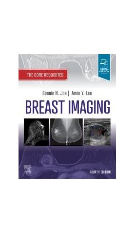
Breast Imaging: The Core Requisites
97.00 ₪
ISBN13
9780323758499
מהדורה
4th edition
הוצאה לאור
Elsevier
עמודים / Pages
526
פורמט
Paperback / softback
תאריך יציאה לאור
13 באוק׳ 2022
Focusing on high-yield information, Breast Imaging: The Core Requisites, 4th Edition emphasizes the basics to help you establish a foundational understanding of breast imaging during rotations, refresh your knowledge of key concepts, and learn strategies to provide "value-added" reports to referring clinicians. This completely rewritten and reorganized edition emphasizes the essential knowledge you need in an easy-to-read format, with thorough updates that cover new imaging modalities, the latest guidelines, and integration of physics information throughout.
-
- Emphasizes the essentials in a templated, quick-reference format that includes numerous outlines, tables, pearls, boxed material, and bulleted content for easy reading, reference, and recall.
- Helps you build and solidify core knowledge to prepare you for clinical practice with critical, up-to-date information on mammography, breast ultrasound, digital breast tomosynthesis, and breast MRIs, as well as special chapters on lymph node evaluation in breast imaging, augmented and reconstructed breast, and special populations in breast imaging.
- Features hundreds of high-quality images, including correlations of ultrasound, mammography, digital breast tomosynthesis and MRI.
- Published as part of the newly reimagined Core Requisites series, an update to the popular Requisites series aimed at radiology trainees and today’s busy clinicians.
- An eBook version is included with purchase. The eBook allows you to access all of the text, figures and references, with the ability to search, customize your content, make notes and highlights, and have content read aloud.
| מהדורה | 4th edition |
|---|---|
| עמודים / Pages | 526 |
| פורמט | Paperback / softback |
| הוצאה לאור | Elsevier |
| תאריך יציאה לאור | 13 באוק׳ 2022 |
| תוכן עניינים |
|
| Author | Bonnie N. Joe, MD, PhD and Amie Y. Lee, MD |













Login and Registration Form