אנו משתמשים ב-Cookies כדי לשפר את החוויה שלך. כדי לקיים ההנחיה החדשה של e-Privacy, עלינו לבקש את הסכמתך להגדיר את ה-Cookies. קבלת מידע נוסף.
1,304.00 ₪
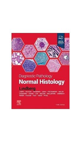
Diagnostic Pathology: Normal Histology
1,304.00 ₪
ISBN13
9780323834148
מהדורה
3rd ed
עמודים / Pages
560
תאריך יציאה לאור
31 באוק׳ 2022
Features more than 2,000 print and online images, including carefully annotated photomicrographs, gross images, electron micrographs, and full-color medical illustrations
This expert volume in the Diagnostic Pathology series is an excellent point-of-care resource for practitioners at all levels of experience and training. Covering all aspects of normal histology of every organ system, it incorporates the most recent scientific and technical knowledge in the field to provide a comprehensive overview of all key issues relevant to today’s practice. Richly illustrated and easy to use, the third edition of Diagnostic Pathology: Normal Histology is a visually stunning, one-stop resource for every practicing pathologist, resident, student, or fellow as an ideal day-to-day reference or as a reliable training resource.
Key Features
- Covers all areas of normal histology, including introductory chapters on electron microscopy, immunohistochemistry and histochemistry, the cell, and the basic organization of tissues
- Includes important updates throughout, covering not only traditional normal histology, but also its morphologic spectrum (variant normal histology) as well as recent advances in immunohistochemistry that expand the spectrum of antigen expression in normal tissues
- Contains new images in over 50% of the chapters, including images of the most common abnormal findings in each organ system, helping provide direct contrast with adjacent normal histology (i.e., what is normal and what is not)
- Provides the at-a-glance information necessary for diagnosis or adequacy evaluation at the time of procedure, using a concise, synoptic writing style
- Features more than 2,100 print and online images, including carefully annotated photomicrographs, gross images, electron micrographs, and full-color medical illustrations to help practicing and in-training pathologists reach a confident diagnosis
- Employs consistently templated chapters, bulleted content, key facts, a variety of test data tables, annotated images, and an extensive index for quick, expert reference at the point of care
- An eBook version is included with purchase. Giving you the power to access all of the text, figures and references, with the ability to search, customize your content, make notes and highlights, and have content read aloud.


Login and Registration Form