אנו משתמשים ב-Cookies כדי לשפר את החוויה שלך. כדי לקיים ההנחיה החדשה של e-Privacy, עלינו לבקש את הסכמתך להגדיר את ה-Cookies. קבלת מידע נוסף.
867.00 ₪
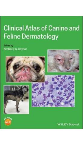
Clinical Atlas of Canine and Feline Dermatology
867.00 ₪
ISBN13
9781119226307
יצא לאור ב
Hoboken
עמודים / Pages
448
פורמט
Hardback
תאריך יציאה לאור
10 בספט׳ 2019
Clinical Atlas of Canine and Feline Dermatology presents more than a thousand high-quality color photographs depicting common dermatologic diseases and conditions, making it easy for clinicians to quickly evaluate and accurately identify clinical dermatologic lesions. Easy-to-use charts of dermatologic diseases provide differential diagnoses and treatments, helping practitioners to quickly find the most common differential diagnoses, perform appropriate diagnostics, and treat their patients.
Written by experienced veterinary dermatologists, the book begins with chapters on essential dermatologic diagnostics and identification and interpretation of skin lesions, featuring pictorial illustrations with commentary of the most common causes. Diagnostic algorithms for pruritus and alopecia simplify the workup of these very common presenting symptoms, and easily referenced tables detail the presentation, diagnosis, and management of hundreds of skin diseases. The book also offers a dermatologic formulary including systemic and topical therapies.
Provides more than 1200 images showing the most encountered dermatologic conditions in dogs and cats
Includes easy-to-interpret charts of differential diagnoses and treatments
Offers diagnostic and treatment algorithms for the most common skin diseases in dogs and cats
Presents details of the presentation, diagnosis, and management of hundreds of skin diseases in tables for quick reference
Features video clips on a companion website demonstrating dermatologic diagnostic techniques, including skin scrapings and cytology, aspiration of skin masses for cytology, and biopsy
Offering fast access to practical information for diagnosing and treating dermatologic disease in small animal practice, Clinical Atlas of Canine and Feline Dermatology is an essential book for any small animal practitioner or veterinary student.
| עמודים / Pages | 448 |
|---|---|
| פורמט | Hardback |
| ISBN10 | 1119226309 |
| יצא לאור ב | Hoboken |
| תאריך יציאה לאור | 10 בספט׳ 2019 |
| תוכן עניינים | List of Contributors Preface Acknowledgement Chapter 1: Dermatology Diagnostics - pictorial description of how to correctly perform and interpret common dermatologic tests; short videos of techniques for skin scrapings, skin and ear cytology and skin biopsy technique are available online A. Skin scrapings B. Cytology - Skin and ear C. Cytology - Mass aspirates D. Trichograms E. Dermatophyte culture technique F. Wood's lamp examination G. Dermatophyte culture medium selection and incubation H. Identification of dermatophytes I. Dermatophyte PCR J. Bacterial culture K. Skin biopsies L. Allergy testing Chapter 2: Dermatology lesions and differential diagnoses - pictorial illustration of different skin lesions with commentary of the most common causes based on lesion number, distribution and location Primary lesions A. Macule/Patch B. Papule/pustule C. Plaque D. Vesicle/bulla E. Wheal F. Nodule G. Cyst Primary or secondary A. Alopecia B. Scale/crust C. Follicular cast D. Comedone E. Pigment change Secondary lesions A. Epidermal collarette B. Scar C. Excoriation D. Erosion E. Ulcer F. Lichenification G. Callus H. Fissure Chapter 3: Lesion location and differentials - pictorial illustrations of common diseases in different body areas A. Face i. Nasal planum ii. Lips/Eyelids iii. Muzzle B. Ears i. Pinnal margin ii. Pinna iii. Outer ear canal C. Feet i. Interdigital ii. Palmar metacarpal/metatarsal iii. Pawpad iv. Nailbed D. Claws E. Perianal/perivulvar F. Tail G. Pressure points H. Trunk - Dorsal and/or lateral I. Inguinal/axillary J. Oral cavity Chapter 4: The pruritic patient - algorithms of the most common diagnoses and diagnostic approach, dog and cat. Chapter 5: The alopecic patient - algorithms of the most common diagnoses and diagnostic approach, dog and cat. Chapter 6: Breed related skin disease - Table of breeds with pictures of unique breed related dermatologic disorders. Chapter 7: Parasitic skin diseases: Table A. Demodex, Sarcoptes, Notoedres, Otodectes, cat fur mite, Cheyletiella, lice, chiggers, hookworm dermatitis, Cuterebra, myiasis, flybite dermatitis. Pelodera dermatitis, dracunculiasis, spider bites, fleas, ticks, chiggers, nematodes Chapter 8: Bacterial, fungal, oomycete and algal infections: Tables and algorithms for workup of recurrent bacterial pyoderma and treatment of generalized dermatophytosis A. Table 8.1: Superficial pyoderma (Impetigo, pyotraumatic dermatitis, intertrigo, mucocutaneous pyoderma, bacterial overgrowth syndrome, bacterial folliculitis) B. Table 8.2: Deep pyoderma (bacterial furunculosis, canine acne, callus furunculosis, acral lick dermatitis, pedal folliculitis/furunculosis, post-grooming furunculosis) C. Table 8.3: Methicillin resistance D. Table 8.4: Underlying causes of canine recurrent pyoderma E. Table 8.5: Commonly used antibiotics for canine pyoderma F. Table 8.6: Topical antibacterial products G. Table 8.7: Subcutaneous bacterial infections (abscess, botryomycosis, cellulitis, necrotizing fasciitis, actinomycosis, nocardiosis, plague L-form infections) H. Table 8.8: Mycobacterial infections I. Table 8.9 Yeast infections (Malassezia, Candida) J. Table 8.10: Dermatophytosis K. Table 8.11: Environmental decontamination in dermatophytosis L. Table 8.12: Deep fungal, oomycete and algal infections (blastomycosis, cryptococcosis, histoplasmosis, Coccidioidomycosis, phaeohyphomycosis, pythiosis, lagenidiosis, zygomycosis, protothecosis) Chapter 9: Viral, rickettsial and protozoal dermatologic diseases: Tables A. Table 9.1: Viral dermatologic diseases (Herpesvirus dermatitis, Calicivirus dermatitis, papillomas, poxvirus, feline sarcoid, feline infectious peritonitis associated dermatitis, canine distemper associated skin lesions) B. Table 9.2: Rickettsial diseases (Rocky Mountain Spotted fever, ehrlichiosis) C. Table 9.3: Protozoal diseases (Leishmania, Toxoplasma) Chapter 10: Allergic skin diseases: Tables and Atopy treatment algorithm A. Table 10.1: Hypersensitivity Disorders, and Treatment of Allergic Skin Diseases B. Table 10.2: Allergy Treatment Toolkit C. Table 10.3: Allergy Testing: Intradermal and Serologic Methods D. Table 10.4: Considerations in Allergen Formulation E. Tables 10.5A and 10.5B: Protocols for Allergen Specific Immunotherapy (ASIT) F. Table 10.6: Performing an Adequate Diagnostic Hypoallergenic Diet Trial G. Table 10.7: Feline Manifestations of Cutaneous Allergy H. Table 10.8: Eosinophilic Granuloma Complex Chapter 11: Autoimmune/immune mediated diseases: Tables and treatment algorithms for canine and feline pemphigus foliaceus A. Table 11.1 Autoimmune/immune mediated diseases (Discoid lupus erythematosus, pemphigus foliaceus, pemphigus vulgaris, vesicular cutaneous lupus erythematosus, mucocutaneous lupus erythematosus, alopecia areata, uveodermatologic syndrome, autoimmune subepidermal blistering diseases (AISBD), vasculitis, post vaccination injection site alopecia, drug eruption, erythema multiforme, toxic epidermal necrolysis, sterile panniculitis, sterile granuloma/pyogranuloma, juvenile cellulitis, plasma cell pododermatitis, symmetric lupoid onychitis, nasal arteritis, metacarpal/metatarsal fistulas, canine sterile neutrophilic dermatitis (Sweet's syndrome), eosinophilic dermatitis (Well's syndrome), superficial suppurative necrolytic dermatitis, systemic lupus erythematosus) B. Table 11.2: Typical glucocorticoid doses for treatment of autoimmune and immune-mediated disorders C. Table 11.3: Oral immunosuppressant or immunomodulatory drugs used as adjunctive treatments of autoimmune/immune-mediated diseases or as primary treatment Chapter 12: Endocrine skin diseases: Tables A. Table 12.1: Canine endocrine alopecia (Hypothyroidism, Cushing's (spontaneous and iatrogenic), Atypical Cushing's, Food-induced Cushing's, topical corticosteroid application, pituitary dwarfism, calcinosis cutis, exogenous estrogen-related alopecia, spontaneous hyperestrogenism, spontaneous hyperandrogenism, tail gland hyperplasia) B. Table 12.2: Trilostane treatment and monitoring C. Table 12.3: Feline endocrine alopecia (hyperthyroidism, hypothyroidism, hyperadrenocorticism, feline acquired skin fragility, diabetes mellitus, acromegaly) Chapter 13: Non-endocrine alopecia: Tables A. Table 13.1: Non-Endocrine, Non-inflammatory Alopecia of Dogs (Post clipping alopecia, traction alopecia, congenital follicular/ectodermal dysplasia, color dilution alopecia, black hair follicular dysplasia, non color, breed-related follicular dysplasia, cyclic flank alopecia, pattern alopecia, follicular lipidosis, Alopecia X, anagen/telogen effluvium) B. Table 13.2: Non-Endocrine, Non-inflammatory Alopecia of Cats (congenital hypotriochosis, hair shaft disorder of Abyssinian cats, pili torti, feline preauricular alopecia, feline pinnal alopecia, feline psychogenic alopecia, mural folliculitis, mucinotic mural folliculitis, pseudopelade, trichorrhexis nodosa, feline paraneoplastic alopecia Chapter 14: Diagnosis and treatment of acute and chronic otitis- detailed discussion and algorithms of the most common diagnoses and diagnostic approach for acute and chronic otitis A. Approach to otitis B. Otoscopic examination C. Choice of otic medications D. Indications for systemic steroid/antibiotic therapy in otitis treatment E. Choice of otic cleansers/flushes F. Educate owners on how to correctly use ear flushes G. Diagnosis and treatment of otitis media H. When to refer for surgery I. Ototoxicity Chapter 15: Metabolic/nutritional/keratinization dermatologic disorders: Table A.Seborrhea (secondary), Vit A responsive dermatosis, sebaceous adenitis, Schnauzer comedo syndrome, nasodigital hyperkeratosis, callus, xeromycteria, ear margin dermatosis, canine acne, feline acne, zinc responsive dermatosis, necrolytic migratory erythema, thymoma induced exfoliative dermatitis, xanthoma, split pawpad disease Chapter 16: Congenital/hereditary skin disorders: Table A. Primary seborrhea, idiopathic facial dermatitis of Persians, ichthyosis, nasal parakeratosis of Labs, dermatomyositis, congenital alopecia, cutaneous asthenia, mucinosis, urticaria pigmentosa, ulcerative dermatitis of Bengal cats, dermoid sinus, acrodermatitis, acral mutilation syndrome, congenital KCS and ichthyosiform dermatosis in CKCS, exfoliative cutaneous lupus erythematosus, epidermolysis bullosa Chapter 17: Pigmentary disorders: Table A. Lentigo, acquired hormone associated hyperpigmentation, acquired post inflammatory hyperpigmentation, vitiligo, nasal hypopigmentation disorders, aurotrichia, "Dalmation bronzing" syndrome Chapter 18: Environmental skin disorders: Table A. Solar dermatitis, burn, radiant heat dermatitis, frostbite, irritant contact dermatitis, grass awns/burrs, post -traumatic alopecia, hygroma, pressure sore Chapter 19: Skin tumors: Table A. Squamous cell carcinoma and squamous cell carcinoma in situ, Bowenoid in situ carcinoma, basal cell carcinoma, sebaceous gland tumors, hair follicular tumors and cysts, cutaneous horns, apocrine gland tumors, apocrine cystomatosis, perianal gland tumors, anal sac tumors, lipoma, infiltrative lipoma, liposarcoma, mast cell tumor, fibroma, dermatofibroma, nodular dermatofibrosis, acrochordon, mammary tumors, hemangioma, hemangiosarcoma, cutaneous progressive angiomatosis, hemangiopericytoma, lymphangioma, lymphangiosarcoma, fibrosarcoma, mammary tumors, cutaneous epitheliotropic lymphoma, cutaneous non-epitheliotropic lymphoma, feline cutaneous lymphocytosis, plasmacytoma, melanocytoma, malignant melanoma, histiocytoma, cutaneous histiocytosis, systemic histiocytosis, feline progressive histiocytosis, cutaneous Langerhans cell histiocytosis, collagenous hamartoma, calcinosis circumscripta, transmissible venereal tumor, feline lung-digit syndrome Chapter 20: Dermatology Formulary: Tables A. Table 20.1: Systemic antibiotics B. Table 20.2: Systemic antifungals C. Table 20.3: Systemic antiviral/antiprotozoal medications D. Table 20.4: Antihistamines E. Table 20.5: Systemic glucocorticoids F. Table 20.6: Immunosuppressive/immunomodulatory drugs G. Table 20.7: Behavior-modifying drugs H. Table 20.8: Antiparasitic medications I. Table 20.9: Nutritional supplements J. Table 20.10: Non-glucocorticoid hormones K. Table 20.11: Topical non-steroidal antipruritic therapies L. Table 20.12: Topical glucocorticoids M. Table 20.13: Topical antimicrobials/otics N. Table 20.14 Topical antiseborrheics O. Table 20.15: Topical immunomodulators and retinoids P. Table 20.16 Topical antiparasitics |


Login and Registration Form