אנו משתמשים ב-Cookies כדי לשפר את החוויה שלך. כדי לקיים ההנחיה החדשה של e-Privacy, עלינו לבקש את הסכמתך להגדיר את ה-Cookies. קבלת מידע נוסף.
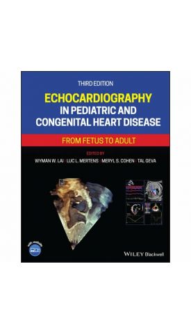
Echocardiography in Pediatric and Congenital Heart Disease: From Fetus to Adult
The new edition of the acclaimed reference text on the most critical tool in pediatric cardiology practice
Echocardiography in Pediatric and Congenital Heart Disease provides comprehensive guidance on the use of non-invasive ultrasound imaging in the diagnosis and treatment of pediatric cardiac conditions. Written by a team of experts from the world’s leading pediatric cardiology centers, this highly-illustrated, full-color reference covers anatomy, pathophysiology, ultrasound physics, laboratory setup, patient preparation and safety, pediatric echocardiogram protocols, quantitative methods of echocardiographic evaluation, and more.
Offering a wealth of additional material on state-of-the-art techniques and technologies in echocardiography, the thoroughly revised third edition features entirely new chapters on examination guidelines and standards, quality improvement in the laboratory, perioperative echocardiography, hemodynamic assessment of the neonate, early fetal echocardiography, and multimodality imaging. This edition offers updated and expanded discussion of the latest advances in echocardiography, particularly those related to speckle tracking and 3D echocardiography. An essential resource for all practitioners, instructors, and trainees in the field, Echocardiography in Pediatric and Congenital Heart Disease:
- Provides up-to-date reference to ultrasound imaging of the hearts of fetuses, children, and adults with both acquired and congenital heart disease
- Covers the echocardiographic examination of congenital cardiovascular abnormalities before, during, and after treatment
- Describes quantitative methods of echocardiographic evaluation, including assessment of diastolic function, right ventricular function and assessment of the post-Fontan patient
- Discusses intraoperative echocardiography, heart disease in pregnancy, and other special techniques and topics
- Includes more than 1200 high-quality color images as well as a companion website with over 600 video clips
Echocardiography in Pediatric and Congenital Heart Disease, Third Edition, remains an essential textbook for cardiac sonographers, pediatric and adult cardiologists, echocardiography nurses and technicians, and adult cardiologists with interest in congenital heart disease.
| מהדורה | 3rd ed |
|---|---|
| עמודים / Pages | 1056 |
| הוצאה לאור | WILEY |
| תאריך יציאה לאור | 23 בדצמ׳ 2021 |
| תוכן עניינים | Preface xii About the Companion Website xiii Part I Introduction to Cardiac Ultrasound Imaging 1 Ultrasound Physics 3 2 Instrumentation, Patient Preparation, and Patient Safety 21 3 Segmental Approach to Congenital Heart Disease 34 4 The Normal Pediatric Echocardiogram 47 Part II Quantitative Methods 5 Structural Measurements and Adjustments for Growth 69 6 Hemodynamic Measurements 87 7 Left Ventricular Systolic Function Assessment 116 8 Diastolic Ventricular Function Assessment 146 9 Right Ventricular Function 168 Part III Anomalies of the Systemic and Pulmonary Veins, Septa, and Atrioventricular Junction 10 Pulmonary Venous Anomalies 189 11 Systemic Venous Anomalies 213 12 Anomalies of the Atrial Septum 228 13 Ventricular Septal Defects 247 14 Ebstein Anomaly, Tricuspid Valve Dysplasia, and Right Atrial Anomalies 267 15 Mitral Valve and Left Atrial Anomalies 280 16 Common Atrioventricular Canal Defects 297 Part IV Anomalies of the Ventriculoarterial Junction and Great Arteries 17 Anomalies of the Right Ventricular Outflow Tract and Pulmonary Valve 321 18 Pulmonary Atresia with Intact Ventricular Septum 340 19 Abnormalities of the Ductus Arteriosus and Pulmonary Arteries 360 20 Anomalies of the Left Ventricular Outflow Tract and Aortic Valve 382 21 Hypoplastic Left Heart Syndrome 405 22 Aortic Arch Anomalies: Coarctation of the Aorta and Interrupted Aortic Arch 430 23 Tetralogy of Fallot 463 24 Truncus Arteriosus and Aortopulmonary Window 492 25 Transposition of the Great Arteries 508 26 Double‐Outlet Ventricle 534 27 Physiologically “Corrected” Transposition of the Great Arteries 560 Part V Miscellaneous Cardiovascular Lesions 28 Hearts with Functional Single Ventricle, Superior‐Inferior Ventricles, and Crisscross Heart 585 29 Echocardiographic Assessment of Functional Single Ventricles after the Fontan Operation 616 30 Cardiac Malposition and Heterotaxy Syndrome 635 31 Congenital Anomalies of the Coronary Arteries 658 32 Vascular Rings and Slings 685 33 Connective Tissue Disorders 700 34 Cardiac Masses 728 Part VI Anomalies of the Ventricular Myocardium 35 Dilated Cardiomyopathy, Myocarditis, and Heart Transplantation 747 36 Hypertrophic Cardiomyopathy 772 37 Restrictive Cardiomyopathy and Pericardial Diseases 794 38 Other Anomalies of the Ventricular Myocardium 822 Part VII Acquired Pediatric Heart Disease 39 Kawasaki Disease 845 40 Acute Rheumatic Fever and Rheumatic Heart Disease 856 41 Infective Endocarditis 870 Part VIII Special Techniques and Topics 42 Transesophageal and Intraoperative Echocardiography 885 43 Pregnancy and Heart Disease 904 44 Fetal Echocardiography 924 45 Echocardiographic Assessment of the Transitional Circulation 964 46 Echocardiographic Assessment of Pulmonary Hypertension 992 47 The Role of Multimodality Cardiovascular Imaging 1011 Index 1031 |
| Author | Wyman W. Lai (Editor), Luc L. Mertens (Editor), Meryl S. Cohen (Editor), Tal Geva (Editor) |


Login and Registration Form